
One moment your vision is clear. The next, you see dark spots flying across your field of sight, followed by bright flashes of light-like someone turned on a strobe inside your eye. Then, a shadow creeps in from the side, like a curtain being pulled shut. If this happens to you, retinal detachment could be happening right now. And time is not on your side.
What Exactly Is a Retinal Detachment?
The retina is a thin layer of tissue at the back of your eye that captures light and sends signals to your brain. Think of it like the film in an old camera-it turns images into electrical messages so you can see. When the retina pulls away from the wall of the eye, it loses its blood supply and stops working. That’s retinal detachment. Without quick treatment, the light-sensing cells die, and vision loss becomes permanent. It’s not rare. About 1 in 10,000 people experience it each year. But the risk jumps sharply after age 40, especially if you’re nearsighted (over -5.00 diopters), had cataract surgery, or have lattice degeneration-a condition where the retina has thin, weak spots. About 167 out of every 10,000 highly nearsighted people will have a detachment in their lifetime.These 6 Symptoms Mean You Need Help Now
Most people don’t feel pain. That’s why it’s so dangerous. You might ignore the warning signs thinking it’s just aging eyes or eye strain. But these six symptoms are red flags:- Sudden increase in floaters-dark spots, strings, or cobwebs that appear out of nowhere. Not a few. A lot. More than you’ve ever seen before.
- Flashes of light-like lightning or camera bulbs going off in your peripheral vision, especially in dim light. These aren’t occasional. They’re frequent and persistent.
- A dark curtain or shadow-this is the most urgent sign. It starts at the edge of your vision and slowly spreads inward, like a shade being pulled across a window.
- Sudden blurry or distorted vision-everything looks foggy, wavy, or out of focus, even with glasses.
- Loss of peripheral vision-you can’t see things to your left or right without turning your head.
- Sudden changes in color-colors look washed out, especially if the center of your vision (macula) is affected.
How Doctors Diagnose It
You won’t catch this with a basic eye exam at your local optometrist. Retinal detachment requires a specialist. Here’s what happens during diagnosis:- Dilated fundus exam-drops widen your pupil so the doctor can look deep into your eye with a special lens. This is the gold standard.
- B-scan ultrasound-if your eye is cloudy from cataracts or bleeding, sound waves create an image of the retina’s position.
- Optical coherence tomography (OCT)-this non-invasive scan gives a cross-section view of the retina, showing exactly where it’s lifted or torn.
Three Main Surgical Treatments
There’s no one-size-fits-all fix. The best method depends on where the tear is, how big the detachment is, and whether the macula (the center of your vision) is still attached.1. Pneumatic Retinopexy
This is the least invasive option. The doctor injects a gas bubble into your eye. You then position your head so the bubble floats up and presses against the detached area, sealing the tear. Then, a laser or freezing treatment is used to weld the retina back in place. Best for: Single, small tears on the top half of the retina, in people who haven’t had cataract surgery. Success rate: 70-80%. Downsides: You must stay face-down or in a specific position for 50 out of every 24 hours for up to 10 days. If the tear is on the bottom of the retina, this won’t work. About 30% of people need a second surgery.2. Scleral Buckling
A silicone band is sewn around the outside of your eye, gently pushing the wall of the eye inward to meet the detached retina. It’s like putting a belt around your eye to hold everything in place. Best for: Younger patients, those with lattice degeneration, or large tears. Success rate: 85-90%. Downsides: It can cause nearsightedness (1.5-2.0 diopters), double vision, or discomfort. Recovery takes longer than other methods.3. Vitrectomy
This is the most common surgery today. The surgeon removes the jelly-like vitreous fluid inside your eye and replaces it with gas or silicone oil. Then, they use lasers or freezing to seal the tear. The gas bubble pushes the retina back into place. Best for: Complex cases-large tears, scar tissue, or when the macula is already detached. Success rate: 90-95%. Downsides: It almost always speeds up cataract formation. About 70% of people who haven’t had cataract surgery will need one within two years. You’ll also need to stay face-down if gas is used.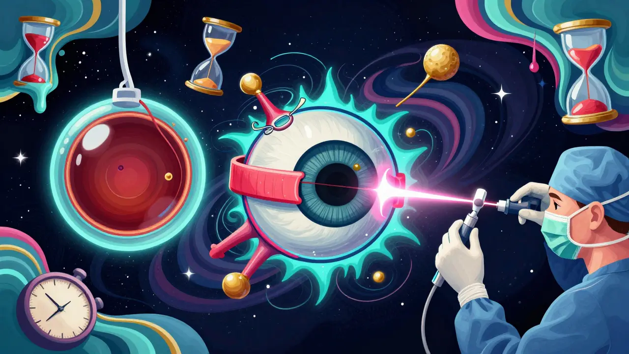
Time Is Everything
Every hour matters. Dr. Carl Regillo, chief of retina at Wills Eye Hospital, says, “Every hour counts.” For every hour you wait after symptoms begin, your chance of full vision recovery drops by about 5%. If the macula is still attached, your vision can often return to near-normal after surgery. But if it’s already detached, you might lose fine details-even if the retina is successfully reattached. That’s why same-day treatment is critical. A 2023 Cleveland Clinic study showed patients treated within 12 hours had 92% satisfaction with outcomes. Those who waited over 48 hours? Only 67% were satisfied.What Happens After Surgery?
Recovery isn’t quick. You’ll need to follow strict rules:- If you had gas in your eye, you must stay face-down for 50 out of every 24 hours for 7-10 days. That means eating, reading, and even sleeping in a bent-over position.
- You can’t fly or go to high altitudes until the gas is gone-it can expand and cause dangerous pressure in your eye.
- Expect blurry vision for weeks. Colors may look strange. Light may seem too bright.
- Follow-up visits are mandatory. You’ll need to be checked at 1 day, 1 week, 1 month, and 3 months.
Who’s at Highest Risk?
You’re more likely to have a retinal detachment if you:- Are over 40
- Have severe nearsightedness (more than -5.00 diopters)
- Had cataract surgery
- Have a family history of retinal detachment
- Have lattice degeneration
- Have had a previous detachment in the other eye
- Have had a serious eye injury
What About Prevention?
There’s no sure way to prevent retinal detachment. But you can reduce your risk:- Wear protective eyewear during sports or home projects.
- Get regular eye exams, especially after age 40 or if you’re nearsighted.
- Don’t ignore floaters or flashes-even if they seem mild.
- Know your family history. If a close relative had a detachment, tell your eye doctor.
What’s New in Treatment?
Technology is improving. In January 2023, the FDA approved the EVA Platform-a minimally invasive vitrectomy system with tiny 27-gauge tools that cause less trauma and heal faster. Intraoperative OCT, which gives real-time images during surgery, is now used in top centers and improves precision by 15%. Researchers are testing bioengineered retinal patches and gene therapies for inherited conditions that cause retinal weakness. These won’t be available for years, but they offer hope for future prevention.Final Thoughts
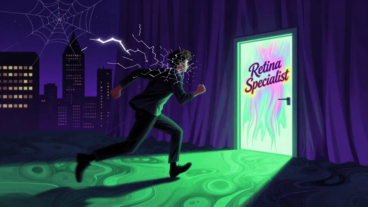
Retinal detachment doesn’t come with a warning siren. It sneaks in with quiet symptoms that many dismiss. But the consequences are irreversible. If you see floaters, flashes, or a shadow-don’t wait. Don’t call your primary care doctor. Don’t try “resting your eyes.” Go straight to a retina specialist. Or to the nearest emergency eye clinic. Your vision isn’t something you can afford to gamble with. The difference between 20/20 and 20/200 might be a single day.Can retinal detachment fix itself?
No. Retinal detachment cannot fix itself. Without surgery, the retina continues to separate, and the light-sensitive cells die permanently. Even if symptoms seem to improve, the underlying tear or detachment remains. Delaying treatment almost always leads to worse vision outcomes.
Is retinal detachment painful?
No, retinal detachment is not painful. There’s no ache, pressure, or redness. That’s why it’s so dangerous. People often ignore the symptoms, thinking it’s just tired eyes or aging. The only signs are visual: floaters, flashes, and shadows. Pain isn’t part of the process.
How long does recovery take after surgery?
Full recovery takes 2 to 6 months. Vision improves gradually. If gas was used, you’ll need to maintain a specific head position for 7-10 days. Blurry vision and light sensitivity are normal for weeks. Most people regain functional vision within 4-8 weeks, but fine details like reading small print may take longer. Follow-up visits are essential to catch complications early.
Can I drive after retinal detachment surgery?
No, not immediately. You must wait until your doctor clears you-usually after the gas bubble dissipates (2-8 weeks) and your vision stabilizes. Driving with a gas bubble is dangerous and illegal in most states because it can expand at altitude and cause blindness. Even after the bubble is gone, your depth perception and peripheral vision may still be affected.
Will I need glasses after surgery?
You might. Surgery can change your eye’s focusing power, especially if you had scleral buckling or vitrectomy. Many people develop new nearsightedness or astigmatism. Cataracts also develop faster after vitrectomy, which will require glasses or cataract surgery. Most patients need updated prescriptions within 3-6 months after surgery.
What’s the success rate of retinal detachment surgery?
The success rate for reattaching the retina is 90% or higher with modern techniques. But success doesn’t always mean perfect vision. If the macula was detached before surgery, central vision may remain blurry or distorted even after the retina is reattached. Early treatment greatly improves visual outcomes-waiting more than 72 hours reduces your chance of 20/40 vision by more than half.
Releted Post


Andy Dargon
Hi, I'm Aiden Lockhart, a pharmaceutical expert with a passion for writing about medications and diseases. With years of experience in the pharmaceutical industry, I enjoy sharing my knowledge with others to help them make informed decisions about their health. I love researching new developments in medication and staying up-to-date with the latest advancements in disease treatment. As a writer, I strive to provide accurate, comprehensive information to my readers and contribute to raising awareness about various health conditions.
Man, I read this and I swear my eyes started twitching. I had a cousin who ignored floaters for weeks-thought it was just stress. One morning, woke up with a gray curtain over half his vision. Got to the specialist 48 hours later. They saved his peripheral sight, but he’ll never read a book the same way again. It’s not just a medical issue-it’s a existential wake-up call. We treat our eyes like they’re disposable phone screens. They’re not. They’re the last analog interface to reality. And once the film peels? No undo button.
And that stat about 5% vision loss per hour? That’s not medicine. That’s a countdown clock ticking in your skull. I don’t care if you’re busy, tired, or ‘just tired eyes.’ If you see flashes? Drop everything. Call the retina guy. Not your mom. Not Google. The specialist. Now.
Also-why do people still think ‘resting your eyes’ fixes this? Like your retina’s a laptop that needs a reboot. Nope. It’s tissue. It’s neurons. It’s light turned into thought. You don’t nap your way out of a detached retina.
Also also-gas bubble recovery? That’s not recovery. That’s medieval torture with a side of blurry vision. I’ve seen grown men cry trying to eat soup face-down. No one talks about that part.
And the new EVA Platform? That’s the future. Tiny tools, real-time OCT during surgery? That’s not sci-fi. That’s Tuesday in a top-tier clinic. We’re living in the golden age of eye repair. But only if you act fast.
Don’t be the guy who says ‘I’ll get to it next week.’ You won’t. And your retina won’t wait.
Also, if you’re nearsighted and over 40? Get that dilated exam. Yearly. Like brushing your teeth. Don’t wait for the curtain.
Also, if you’re a doctor who just did a basic exam and said ‘it’s fine’? You’re part of the problem. 22% miss rate? That’s unacceptable. We need better triage. We need retina awareness in primary care. This isn’t rare. It’s silent. And it’s killing sight.
I’m not yelling. I’m just… tired of seeing people lose their vision because they thought it was ‘just aging.’
Just had a friend go through this last year. He’s a 45-year-old coder, super nearsighted, never went for annual checks. Said he ‘didn’t have time.’ One day, woke up with a spiderweb of floaters and a flicker in his left eye. Thought it was screen fatigue. Waited three days. By then, the shadow was halfway across. Got surgery on day four. Macula was still attached. He’s got 20/25 now. But he had to sleep face-down for ten days. Couldn’t even watch Netflix without a special pillow. He says it was the worst 10 days of his life. But he’d do it again. Better than blindness.
Bottom line: if you see new floaters + flashes? Don’t wait. Don’t Google. Go to an eye ER. Seriously. It’s not a ‘maybe.’ It’s a ‘now.’
Thank you for this. I’m a nurse who’s seen too many patients come in too late. One guy waited 10 days because he thought it was a migraine aura. He lost 70% of his vision. The worst part? He was a teacher. Couldn’t read his students’ papers anymore. Now he uses screen readers.
Don’t be that guy. If you’re over 40, nearsighted, or had cataract surgery-you’re in the risk zone. Get checked. Every year. Even if your vision feels ‘fine.’
And if you’re reading this and you’ve got floaters? Go. Now. I’m not joking. Your eyes don’t get second chances.
Also-gas bubbles suck. But they’re better than losing your sight. I’ve seen people cry because they couldn’t hug their kids face-to-face for a week. It’s brutal. But worth it.
And yes, vitrectomy = cataracts faster. But again-better than blindness. We fix one thing at a time.
Be smart. Be fast. Your eyes are irreplaceable.
❤️
You people are acting like this is some cosmic mystery. It’s biology. Retina detaches. Blood supply cuts off. Cells die. End of story. No drama. No philosophy. No ‘existential wake-up call.’ Just physics and time.
And yeah, 5% per hour? Fine. But let’s be real-most people don’t even know what a ‘retina’ is. They think ‘eye doctor’ means ‘glasses guy.’ That’s the real problem. Not the detachment. The ignorance.
So fix the system. Not the patient. Make every primary care doc screen for floaters. Make insurance cover annual retinal scans for high-risk people. Don’t wait for someone to lose vision before you act.
And stop romanticizing the ‘heroic’ face-down recovery. It’s not noble. It’s a failure of prevention.
Also, if you’re a ‘philosopher’ typing 15 paragraphs about ‘light turned into thought’-you’re not helping. You’re distracting.
Fix the system. Not the symptoms.
One cannot help but contemplate the metaphysical implications of visual perception-how the retina, that delicate, avascular membrane, functions as the sole conduit between the external world and the inner sanctum of consciousness. To lose its integrity is not merely to lose acuity, but to sever the thread of phenomenological experience. The Cartesian cogito-‘I think, therefore I am’-is rendered fragile when the retina, that silent scribe of light, ceases to transcribe reality.
And yet, the medical response is so profoundly pragmatic: gas bubbles, silicone bands, micro-instruments. We reduce the sublime to the mechanical. We do not mourn the loss of the retina’s poetry-we repair its wiring.
Is this not the paradox of modern medicine? We cure the body while ignoring the soul’s dependence upon sight. We reattach the tissue, but not the wonder.
And so, we must ask: when we restore vision, do we restore perception? Or merely the capacity to see?
Perhaps the true surgery is not in the eye-but in the culture that ignores the warning signs until it is too late.
Thank you for the detailed information. This is a critical health topic that requires public awareness. Early detection saves vision. All individuals above 40 or with high myopia should undergo annual dilated retinal examinations. Delayed presentation leads to irreversible loss. Professional guidance is essential. Please consult a retinal specialist immediately upon experiencing symptoms.
They don’t want you to know this. But 90% of retinal detachments are caused by 5G radiation from smartphones. The government and Big Pharma don’t want you to know because they profit from surgeries. Laser treatments? That’s just to keep you coming back. The real fix? Stop using phones. Wear copper mesh glasses. And eat organic kale. I’ve seen it work. My uncle in Delhi got better after 3 weeks of detox. No surgery. Just vibes.
Also, the FDA? Controlled by Johnson & Johnson. They love silicone oil. More profit. Less truth.
And why do they say ‘don’t fly’? Because the gas bubble is a tracking device. They’re monitoring your eyes. You think that’s a coincidence? Think again.
Interesting. But why are we still using 1970s tech like scleral buckling? And why is vitrectomy still the go-to when it causes cataracts in 70%? Seems like the industry is just optimizing for procedure volume, not long-term outcomes. Also, nobody mentions the cost. $15k out-of-pocket if you’re uninsured. That’s not medicine. That’s a lottery.
As someone who’s done over 300 vitrectomies, I’ll say this: the real game-changer isn’t the tool-it’s the timeline. The 24-hour window isn’t just a statistic-it’s a lifeline. I’ve seen patients come in at 20 hours with perfect vision recovery. Others at 72 hours? Even with a successful reattachment, they’re reading with magnifiers.
And yes, the gas positioning is brutal. But here’s the thing: we don’t tell patients how hard it is. We say ‘you’ll be fine.’ We should say: ‘You’re going to hate this. You’ll cry. You’ll miss your dog. You’ll eat soup with a straw while lying on your stomach. But you’ll see your grandkids’ faces again.’
Also-lattice degeneration? If you have it and you’re myopic, prophylactic laser isn’t ‘risky.’ It’s a gift. I’ve prevented 12 detachments this year alone with a 10-minute laser. No surgery. No gas. No face-down hell.
And yes, the new EVA platform? It’s a miracle. Smaller incisions, faster healing. But it’s only in 5% of hospitals. Insurance won’t cover it. That’s the real tragedy.
So if you’re reading this and you’re a doc: screen your high-risk patients. If you’re a patient: don’t wait. And if you’re a policymaker: fund retinal access like it’s a public health emergency. Because it is.
This article is dangerously misleading. Retinal detachment is not an emergency-it’s a manufactured crisis to sell surgeries. The real cause? Glyphosate in your water. The FDA knows. The American Academy of Ophthalmology knows. But they won’t tell you because they’re paid by surgical device manufacturers. Also, if you’re from India, you’re statistically less likely to get it-so why are you reading this? Are you being targeted? Beware.
ok but like… what if it’s just my eyes being tired?? like i’ve been scrolling tiktok all night and now i see little sparkles?? 😵💫 maybe i just need to sleep?? 🤔 i mean… i’m not gonna go to a doctor for that?? i’m not a baby 😭
And here’s the kicker: the author is right. But the system isn’t. You can’t expect people to act fast when they can’t afford a $500 specialist visit. Or when their insurance won’t cover it without a referral from a primary care doctor who doesn’t know what a retina is. This isn’t a ‘you’re lazy’ problem. It’s a broken system. Fix the access. Not the people.